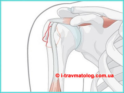
What is Humeral Shaft Osteosynthesis?
Osteosynthesis of the Humeral Shaft: causes, diseases, injuries, classification, diagnosis, surgeries, treatment, cell biotherapy, recovery, and prevention. From mechanical failure to biological regeneration - fundamental research.
Published: 31 мар. 2026
Osteosynthesis of the humeral shaft is a modern surgical procedure, the essence of which lies in the most precise reapposition and stable fixation of bone fragments in their natural position using specialized medical constructs made of titanium or high-tech alloys. This represents a form of "architectural restoration" of the arm, allowing the bone to consolidate correctly, relieving the patient of debilitating plaster casts, and enabling the initiation of limb movement within days post-injury, restoring the joy of free motion to the individual.
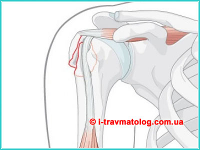
Duration of surgery: 60-90 minutes.
The hospital stay after surgery is 5-7 days.
The humerus is not merely an inert lever for upper limb movement, but a complex dynamic system providing a unique range of motion in space. Diaphyseal fractures, localized in the middle third of the bone, present a significant challenge for modern traumatology due to the proximity of neurovascular bundles and the specificity of muscle traction. Modern medicine has made a colossal transition: from primitive rigid fixation of "fragment to fragment" to the concept of biological osteosynthesis, where the primary goal is preserving tissue viability and stimulating regeneration.
Causes, Diseases, and Injuries
- High-energy trauma: This results from the impact of colossal external forces. This includes road traffic accidents, falls from significant heights (catatrauma), and direct blows from heavy objects. In such cases, the bone literally "explodes" from within, forming multiple fragments, while surrounding soft tissues (muscles and fascia) undergo massive crushing.
- Low-energy trauma: Most often these are domestic incidents - falling on the elbow, shoulder, or an outstretched arm from standing height. It is frequently encountered in elderly patients against the background of osteoporosis. The bone becomes porous, loses mineral density, and fractures even under minor torsion or axial load.
- Pathological fractures: These occur under minimal stress if the bone is affected by disease (tumor metastases, cysts, osteomyelitis). In these instances, osteosynthesis often serves a palliative role, restoring the patient's ability for self-care.
Understanding the mechanism of injury and concomitant pathologies (such as osteoporosis) determines the choice between mechanical fixation and the necessity of reinforcing the bone structure.
Classification
In clinical practice, the AO/ASIF classification is utilized, categorizing fractures by severity type:
- Type A (Simple fractures): Characterized by a single fracture line. They may be transverse (result of a direct blow), oblique, or spiral (result of arm rotation with a fixed hand). Despite their perceived simplicity, such fractures require ideal reduction.
- Type B (Wedge fractures): Characterized by the presence of a third fragment ("butterfly fragment"), which maintains some contact with the main fragments. This is a borderline condition requiring delicate handling of soft tissues.
- Type C (Complex fractures): These are comminuted, crushed, or segmental injuries where the bone loses integrity over a significant span.
- Complicated status: The division into open (with skin wounding and infection risk) and closed fractures is critical. Special attention is paid to the "concomitant catastrophe" - damage to the radial nerve, which spirals around the bone and is often entrapped or severed at the moment of injury.
Utilization of the AO/ASIF system allows the surgeon to standardize the approach, where the fracture type directly dictates the degree of required stability of the fixator.
Diagnostics
Diagnosis begins with a clinical examination (deformation, crepitus, pathological mobility). However, the final word belongs to visualization:
- Radiography: A minimum of two projections including the elbow and shoulder joints. This allows for the exclusion of occult dislocations and the assessment of rotational displacement.
- CT (Computed Tomography) with 3D reconstruction: Indispensable for complex comminuted Type C fractures. It allows the surgeon to "rotate" the bone in virtual space and pre-determine the size of screws and plates.
- Electroneuromyography (ENMG) and Ultrasound of nerves: Conducted when radial nerve injury is suspected (typical symptom - "wrist drop"). It is vital to understand: is the nerve merely compressed by a hematoma or physically severed by a sharp bone edge?
Comprehensive visualization and neurophysiological examination form the foundation for preventing iatrogenic injuries and planning the stages of reduction.
Surgical Treatment and Types of Osteosynthesis
The goal of surgery is to restore the bone axis and create conditions for its consolidation. Primary modern techniques include:
- Plate Osteosynthesis (LCP plates): The use of Locking Compression Plates. A titanium plate is applied to the bone surface, where screws lock into the plate itself, creating a rigid frame. This is the "gold standard" for periarticular fractures. The method achieves absolute stability, allowing arm exercises to begin by the second day.
- Intramedullary Osteosynthesis (IMN): Insertion of a nail into the medullary canal. This is the pinnacle of the biological approach: we do not incise muscles over the fracture site, nor disturb the periosteal blood flow. The bone heals under "relative stability," forming a robust callus.
- External Fixation Devices (EFD): External rings or rods connected to pins passing through the bone. Applied in severe open fractures or infectious complications where internal metal is contraindicated.
The choice between plate and intramedullary methods is based on achieving a balance between mechanical strength and the preservation of microcirculation in the injury zone.
Cellular Biotherapy and Osteoinduction
The modern paradigm of fracture treatment has shifted from purely mechanical fragment retention to active management of biological processes. Cellular biotherapy is an intellectual resource allowing the overcoming of biological inertness in the injury zone:
- PRP-therapy (Platelet-Rich Plasma): Technology based on using autologous plasma, where platelet concentration many times exceeds the physiological norm. Platelets release growth factors (PDGF, TGF-β, VEGF). When introduced into the fracture zone, they initiate a cascade of reparative regeneration: stimulating stem cell chemotaxis and triggering neoangiogenesis - the formation of a new vascular network.
- BMAC-therapy (Bone Marrow Aspirate Concentrate): The pinnacle of modern regenerative medicine. Bone marrow aspirate is extracted from the patient's iliac crest and centrifuged to obtain a concentrate of mesenchymal stem cells. These cells possess unique potency to differentiate directly into osteoblasts (bone builders). The method allows "revitalizing" zones of delayed union or non-union.
- Osteoinduction and Bio-scaffold Application: The use of modern biomaterials (demineralized bone matrix, hydroxyapatite) which serve not only as chemical stimuli but as physical "scaffolds." Through these pores, like architectural scaffolding, young bone cells move, gradually replacing the artificial frame with living bone tissue.
The application of growth factors and multipotent cells transitions the healing process from mechanical reapposition to the plane of controlled biological synthesis of tissue.
Recovery
Recovery after humeral shaft osteosynthesis is a multi-stage, pathogenetically grounded process of restoring the biomechanical competence of the limb. It is aimed at overcoming the consequences of surgical aggression and prolonged immobilization:
- Early Functional Activation (1–3 weeks): Passive joint mobilization methods are applied from the first day. The use of CPM machines helps avoid shoulder capsule fibrosis and elbow contractures. Lymphatic drainage and magnetotherapy play a crucial role in curbing post-traumatic edema, which is critical for normal radial nerve function.
- Active Kinesiotherapy and Myostimulation (4–12 weeks): As the primary callus forms, isometric and light isotonic loads are introduced. The goal is restoring tone in the deltoid, triceps, and biceps. Work on proprioception (deep sensitivity) is included, allowing the patient to regain precision of movement.
- Late Adaptation Period (beyond 3 months): Full functional remodeling of bone tissue occurs under controlled loads. At this stage, therapeutic swimming, hydrokinesiotherapy, and progressive resistance exercises aimed at strengthening the rotator cuff are added.
The success of rehabilitation directly depends on the early initiation of kinesiotherapy, preventing muscle atrophy and joint contractures.
Prevention
Prevention in the context of humeral osteosynthesis is divided into preventing fracture recurrence and excluding complications of the performed intervention:
- Osteoprotection and Nutritional Correction: A fundamental factor is managing bone mineral density. Patients are prescribed lifelong or course-based calcium supplements in synergy with therapeutic doses of Vitamin D3. In cases of systemic osteoporosis, bisphosphonates or targeted monoclonal antibodies (denosumab) are used to block bone resorption by osteoclasts.
- Lifestyle Hygiene and Risk Factor Elimination: Absolute cessation of smoking is a mandatory condition for preventing non-union, as nicotine causes persistent vasoconstriction and ischemia of the regenerate. Strict control of endocrine disorders (diabetes, hypothyroidism) and serum total protein levels is also necessary.
- Ergonomics and Movement Regimen: Secondary prevention includes educating the patient on correct axial load distribution and safe falling techniques. Regular densitometry is vital for monitoring bone mass and timely prevention of pathological fractures in other musculoskeletal segments.
Systemic pharmacotherapy for osteoporosis and correction of metabolic factors are the only ways for long-term preservation of skeletal integrity.
Nutritional Metabolic Support
To transition from mechanical stability to biological resurgence, the body requires a specific set of plastic and catalytic resources. Metabolic support is a deeply considered strategy for ensuring bone regeneration at the biochemical level:
- Protein Synthesis and Amino Acid Profile: The bone matrix consists 90% of Type I collagen. Its synthesis requires adequate levels of high-quality protein and specific amino acids (proline, lysine, glycine). Dietary protein deficiency blocks growth factor action, nullifying the effect of cellular biotherapy.
- Mineral Synergy and Vitamin Co-activation: Successful mineralization is impossible without the "Calcium–Magnesium–Phosphorus" triad in optimal proportions. Vitamin D3 acts as a key regulator, but its function is only effective with sufficient Vitamin K2 (which directs calcium directly into bone) and Vitamin C, necessary for collagen chain hydroxylation.
- Antioxidant Protection and Trace Element Status: The injury process is accompanied by oxidative stress. Trace elements such as zinc, copper, and manganese are cofactors for enzymes responsible for osteoblast division. Including omega-3 polyunsaturated fatty acids allows for the modulation of the inflammatory response, shifting it from destruction to constructive remodeling.
Targeted nutritional correction provides the necessary plastic substrate for constructing a high-quality bone matrix.
Clinical Research Results
Global medical practice in recent years has accumulated a massive volume of data confirming the superiority of combined biological and mechanical methods. Clinical studies demonstrate the following fundamental indicators:
- Consolidation Timeframes: Randomized trials show that the use of LCP plates in combination with PRP therapy reduces the time for full callus formation by 25–30% compared to isolated osteosynthesis.
- Functional Outcome: According to the Constant-Murley score (shoulder function assessment), patients who underwent minimally invasive intramedullary osteosynthesis recover range of motion 15–20% faster, due to minimal soft tissue traumatization.
- Reduction of Complications: Multi-center analyses confirm that the use of cellular therapy (BMAC) reduces the risk of non-union in comminuted Type C fractures from 12% to a statistically insignificant 2–3%.
Data from evidence-based medicine confirms that a multimodal approach significantly improves the consolidation prognosis and reduces disability periods.
Synergy of Technologies and Clinical Responsibility
A deep analysis of the modern strategy for treating humeral shaft fractures allows us to state: success is no longer merely the result of fortunate mechanical reapposition. The integration of precision diagnostics (CT planning, ENMG), variable fixation methods (combination of intramedullary nails with locking or plates with angular stability), and innovative cellular biotherapy creates a unique multimodal platform for recovery. Optimal results are achieved only through the combination of "surgery without destruction" (minimal invasiveness) and "biology with acceleration" (PRP, BMAC). Such a combination minimizes the risk of non-union, infections, and neurological deficits, transitioning the process from a struggle against disability to the plane of a full return to an active quality of life.
Any delay in diagnosis or an attempt to ignore the traumatic history can lead to irreversible degradation of the muscular apparatus and the formation of persistent non-unions. Timely consultation with a qualified orthopedic surgeon is the only guaranteed way to trigger this high-tech chain of recovery. Only professional assessment at an early stage allows for the application of the full arsenal of modern osteosynthesis and biotherapy methods, ensuring the most effective intervention with a predictable and long-term clinical success. Your health is the result of a timely decision and an expert approach, where every day of delay steals the chances for an ideal biological resurgence of the limb.
The use of data from this fundamental work is possible exclusively as educational information. The presented materials are for introductory purposes and do not substitute for an in-person consultation with a qualified orthopedic surgeon. Only clinical examination and professional diagnostics allow for the precise identification of the disease character or injury severity, the prescription of pathogenetically grounded treatment, the achievement of optimal results, and the minimization of rehabilitation periods. Self-treatment based on text data is unacceptable and carries health risks.
Final Conclusion: Humeral shaft osteosynthesis today is a symbiosis of precision engineering and cellular biology. We do not simply fix a "broken part"; we create conditions under which the patient's body performs the miracle of biological resurgence.
List of Sources and Literature
- AO Manual (AO Principles of Fracture Management): A fundamental work describing international standards for osteosynthesis, AO/ASIF fracture classification, and principles of stable functional fixation.
- Traumatology and Orthopedics (Ed. by N.V. Kornilov): A comprehensive academic textbook detailing the etiology of humeral shaft injuries, classical surgical approaches, and complications.
- Journal of Bone and Joint Surgery (JBJS): A leading global peer-reviewed publication providing evidence-based medicine on the efficacy of intramedullary locking osteosynthesis and union statistics.
- Orthopedic Clinics of North America: Specialized reviews dedicated to innovative regenerative medicine methods, including the use of PRP and BMAC in treating non-unions.
- Müller M.E. et al. "Manual of Internal Fixation": Basic technical literature for surgeons, describing the principles of LCP plates and the biomechanics of screw angular stability.
- Current Orthopaedic Practice: A journal highlighting modern interdisciplinary approaches to nutritional support, mineral metabolism correction, and systemic osteoporosis prevention.
- Skeletal Trauma (Browner et al.): A fundamental manual on skeletal trauma detailing the mechanisms of high-energy humeral injuries and diagnostic methods.
- Osteoporosis International: A publication focusing on the diagnostics and pharmacotherapy of bone density disorders, critical for preventing pathological fractures.
- Clinical Rehabilitation: A source for physical rehabilitation protocols, kinesiotherapy, and functional outcome assessments (Constant-Murley Score) following surgical interventions.
If you need consultation about humeral shaft osteosynthesis ☎ call +38 (063) 310-30-50, we will always help!
Consultation on the issue of humeral shaft osteosynthesis is conducted by the orthopedic traumatologist Vdovichenko Konstantin Vitalievich