Hip Arthroscopy
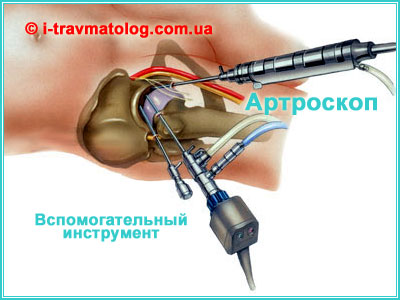
Hip arthroscopy (HA) - is a modern, minimally invasive surgical procedure that allows the surgeon to diagnose and treat problems inside or around the joint without a large incision.
Duration of surgery: 90 minutes.
Hospital stay after surgery: 3-5 days.
Diagnostic hip arthroscopy
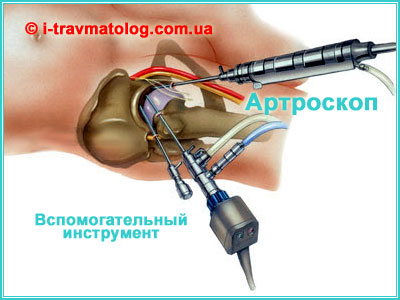
Diagnostic hip arthroscopy is a procedure that allows examination of the space between the femoral head and the hip socket, as well as the rest of the hip. The surgery is performed under general or regional anesthesia. Typically, 3 to 5 small incisions are made to access the hip joint. Arthroscopy is performed with the leg extended to facilitate access to the space between the femoral head and the hip socket. This can be done in the lateral or supine position. We perform hip arthroscopy in the supine position. The surgery is controlled using an image intensifier (a mobile X-ray machine for live imaging), which ensures greater safety during surgery.
Indications for hip arthroscopy:
- the need to remove intra-articular bodies;
- trauma to the labrum in the acetabulum;
- damage to articular cartilage and osteoarthritis;
- development of avascular necrosis of the femoral heads;
- chronic synovitis;
- ruptures of the round ligament;
- septic arthritis;
- instability joints;
- complications after surgical interventions.
1. Indications for Procedure Hip Arthroscopy
Arthroscopy is indicated for both diagnostic and therapeutic purposes, especially when conservative treatment has failed.
Diagnostic Purposes Hip Arthroscopy:
- Identifying the source of pain (when MRI, CT, or ultrasound are inconclusive).
- Assessing the condition of the articular cartilage, labrum, and ligaments.
- Preparation for total hip replacement (analysis of the intra-articular cavity status).
- Biopsy (taking a tissue sample for analysis).
Therapeutic Purposes Hip Arthroscopy:
- Addressing Femoroacetabular Impingement (FAI) - correction of bone overgrowth (osteoplasty) on the femoral head or acetabulum.
- Repair/suture or resection (removal) of a damaged labrum.
- Removal of loose intra-articular bodies (cartilage or bone fragments, known as "joint mice").
- Treatment of articular cartilage injuries (microfracture, debridement, removal of necrotic areas).
- Synovectomy (removal of the inflamed synovial membrane) for chronic synovitis or septic arthritis.
- Repair of damaged ligaments (e.g., the ligamentum teres).
2. How the Surgery is Performed
- Anesthesia: General or spinal (epidural) anesthesia is typically used.
- Positioning: The patient is positioned on a specialized orthopedic traction table.
- Traction: To create working space in the narrow hip joint, controlled traction of the operated limb is applied.
- Access: The surgeon makes 2-4 small punctures (incisions) approximately 5-10 mm long.
- Instrument Insertion:The following are inserted through the punctures:
- Arthroscope: A thin tube with a camera, light source, and irrigation system (to deliver fluid to flush the joint and improve visibility). The image is displayed on a monitor.
- Microsurgical Instruments: Specialized tools for performing therapeutic maneuvers (resection, suturing, shaving/debridement).
- Maneuvers: The surgeon performs the necessary diagnostic or therapeutic procedures under visual control.
- Conclusion: After the procedures are complete, the instruments are removed. The punctures are closed with sutures or sterile dressings.
3. Contraindications
Despite its minimal invasiveness, arthroscopy has a number of contraindications:
- Ankylosis (complete fusion of joint surfaces, lack of mobility).
- Severe osteoarthritis/coxarthrosis of late stages (significant cartilage destruction, where total joint replacement is indicated).
- Active purulent/septic process in the joint or surrounding skin.
- Severe obesity, which may complicate access to the joint.
4. Rehabilitation After Arthroscopy
Rehabilitation - is a critically important stage that largely determines the success of the surgery.
Early Postoperative Stage:
Duration: First 1–2 weeks
Key Points:
- Pain Management (NSAIDs).
- Thrombosis Prevention (anticoagulants).
- Ice application to the joint area.
- Controlled Weight-Bearing on the operated leg using crutches (usually partial or non-weight-bearing, depending on the extent of the intervention).
- Physical Therapy (light movements and isometric exercises, sitting).
Restorative Stage:
Duration: 2-6 weeks
Key Points:
- Gradual increase in weight-bearing (weaning off crutches under physician control).
- Start of active physiotherapy: developing range of motion, strengthening hip and gluteal muscles.
Functional Stage:
Duration: 6 weeks – 3–6 months
Key Points:
- Full restoration of strength and endurance.
- Increasing exercise difficulty: stationary bike, swimming, pool exercises.
- Return to daily activities.
- Return to high-impact sports (e.g., running) is possible no earlier than 3–6 months and only with the doctor's permission.
Important: The exact timeline and rehabilitation program are always individual and depend on the type of maneuver performed (e.g., labral repair requires a longer period of restricted weight-bearing than simple loose body removal). Rehabilitation is supervised by a physical therapist or rehabilitation specialist.