What is Radial Head Replacement?
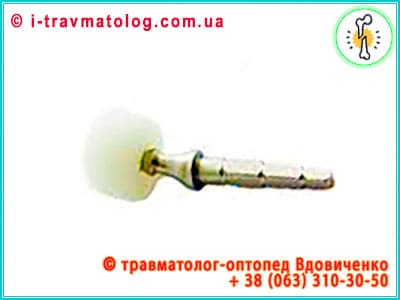
Radial head replacement is a high-tech surgical procedure involving the replacement of a destroyed or irreversibly damaged bone epiphysis with an artificial implant. This field of elbow surgery has evolved from simple metal "plugs" to biomechanically precise systems integrated into the concept of regenerative medicine.
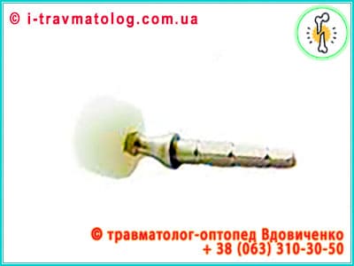
Arthroplasty represents the final stage in the evolution of elbow surgery, transforming a destructive process into a controlled biomechanical reconstruction.
Why Does the Joint Fail?
The primary cause is high-energy trauma. The radial head serves as a key stabilizer of the elbow joint, providing resistance to valgus stress and longitudinal stability of the forearm. Its injury is rarely isolated and is frequently accompanied by ligamentous ruptures.
- Mason Type III and IV Fractures: Critical comminution of the head into three or more fragments, which cannot be restored via open reduction and internal fixation (ORIF) due to the risk of aseptic necrosis or bone mass deficit.
- Elbow Malalignment and Post-traumatic Osteoarthritis: Malunited fractures or chronic instability lead to degenerative destruction of the hyaline cartilage of the humeral capitellum, causing persistent pain and functional limitation.
- Osteonecrosis (Preiser's disease in the context of the radius): Avascular death of bone tissue due to profound microcirculation impairment, leading to the collapse of the articular surface.
- Rheumatoid Arthritis and Systemic Arthropathies: Aggressive synovial inflammation causes bone erosion and destruction of the annular ligament, requiring radical replacement of the articular end to restore biomechanics.
- Congenital Deformities and Neoplastic Processes: Conditions requiring resection of the proximal radius with immediate deficit replenishment to prevent proximal migration of the bone (proximal shift).
Understanding the etiology allows for the identification of high-energy trauma and systemic degeneration as the main factors dictating the need for radical bone deficit replenishment.
Classification
Surgical strategy is based on a deep analysis of injury morphology. The primary tool remains the Mason classification, supplemented by modern understanding of associated instability:
- Type I (Minor Injuries): Marginal fractures without displacement or with displacement of less than 2 mm. Treatment is predominantly conservative with early functional loading.
- Type II (Displaced Marginal Fractures): Fragment displacement greater than 2 mm with preservation of more than 70% of the articular surface area. Internal fixation with microscrews may be performed.
- Type III (Comminuted Fractures): Complete destruction of the head and neck. Reduction is impractical due to the high risk of non-union. This is the primary indication for replacement, especially in the presence of medial collateral ligament injury.
- Type IV (Terrible Triad or Dislocation): Radial head fracture combined with posterior or lateral dislocation of the forearm bones. This condition is characterized by global elbow instability (Hotchkiss modification), where the prosthesis acts as a rigid spacer holding the joint against recurrent dislocation.
The Mason classification serves as the basic algorithm, where Type III and Type IV determine the inevitability of moving from joint-preserving synthesis to arthroplasty.
Diagnostics
The modern "From Visualization to Regeneration" protocol begins with precision multimodal assessment of the damaged segment's condition:
- Radiography in Standard and Special Projections: AP, lateral, and oblique views (Greenspan view) allow for assessment of the degree of displacement and coronoid process involvement.
- CT with Multiplanar 3D Reconstruction: The "gold standard," allowing the surgeon to perform a virtual "assembly" of the joint, assess the size of the defect, and select the prosthesis diameter (from 18 to 24 mm and larger).
- MRI (Magnetic Resonance Imaging): Critically important for assessing the soft tissue component. The integrity of the annular ligament, lateral ulnar collateral ligament (LUCL) complex, and tendons is investigated. Without adequate soft tissue status, even an ideal prosthesis will be unstable.
- Ultrasound (US): Used dynamically to assess the condition of surrounding muscles and detect hematomas in the early post-traumatic period.
Comprehensive diagnostics using 3D reconstruction and MRI guarantee the accuracy of implant sizing and assessment of the soft tissue envelope.
Differential Diagnosis and Identification of "Red Flags"
A critical stage of the examination is the differentiation between primary radial head injury and associated pathologies capable of radically changing the surgical outcome. Detailed differentiation includes:
- Posterior Interosseous Nerve (PIN) Syndrome: It is necessary to distinguish finger extensor palsy caused by direct nerve injury from reflex muscle inhibition due to pain. A "red flag" is the isolated inability to extend at the metacarpophalangeal joints with preserved wrist extension (due to the radial extensors).
- Essex-Lopresti Lesion: This is a critical diagnostic trap. A radial head fracture is combined with an interosseous membrane rupture and dislocation at the distal radioulnar joint (DRUJ). A "red flag" is wrist pain upon palpation following elbow trauma. In this case, head resection without replacement will lead to catastrophic proximal migration of the radius.
- Forearm Compartment Syndrome: High-energy Mason IV fractures are associated with a risk of critical subfascial pressure elevation. "Red flags" include increasing pain unresponsive to analgesics, soft tissue tension, and paresthesias (the "5P" rule).
- Septic Arthritis: In cases of chronic pain or pathological fractures, a septic process must be excluded. A combination of fever, local hyperthermia, and elevated C-reactive protein levels requires joint aspiration before planning replacement.
- Heterotopic Ossification (HO): In the early stages, it can mimic contracture. It is important to differentiate mechanical blocks (prosthesis displacement) from biological blocks (extra-skeletal bone formation), determined by CT data 4-6 weeks after injury.
Timely identification of distal instability (Essex-Lopresti) and neurological deficits is key to preventing irreversible dysfunction of the entire upper limb.
Surgical Approaches and Neural Structure Safety
Implantation success directly depends on the choice of surgical approach, which must provide adequate visualization with minimal soft tissue trauma and protection of critically important nerves.
- Kocher Approach (between the anconeus and the extensor carpi ulnaris): The most common. It provides a direct path to the lateral department of the joint and the annular ligament. The main advantage is the ability to safely visualize the radial head and neck. However, the surgeon must remember the proximity of the posterior interosseous nerve (a branch of the radial nerve), which wraps around the radial neck.
- Kaplan Approach (through the extensor carpi radialis longus and the extensor digitorum communis): Located more anteriorly. This approach minimizes the risk of damage to the lateral collateral ligament complex but requires extreme caution as the posterior interosseous nerve passes just millimeters from the manipulation zone.
- Intraoperative Neuroprotection: To prevent neuropathy, a forearm supination technique is used, which moves the nerve away from the neck resection zone. The use of blunt retractors and the avoidance of aggressive soft tissue stretching are also critically important.
- Ligamentous Apparatus Restoration: Following prosthesis installation, a mandatory stage is the precision reconstruction of the annular ligament and the lateral ulnar collateral ligament (LUCL) using anchor fixators or transosseous sutures. Without this step, even the most advanced implant will be prone to subluxation.
The choice between Kocher and Kaplan approaches requires a balance between visualization and neuroprotection of the posterior interosseous nerve.
Surgical Treatment and Prosthesis Types
Implant choice depends on anatomical features and the degree of ligamentous destruction. Modern systems are classified by fixation method and biomechanics:
- Monoblock (Rigid) Prostheses: Manufactured from titanium or cobalt-chromium alloy. They offer high reliability; however, inaccurate positioning can lead to hyper-pressure and wear of the humeral capitellum cartilage (the "erosion effect").
- Modular Systems: Consist of independent components (head, neck, stem). This allows the surgeon to intraoperatively adjust the prosthesis height, restoring the "length" of the radius with millimeter precision, which is critical for ligamentous tension.
- Bipolar (Floating) Prostheses: Possess an additional internal degree of freedom. The head can perform tilting movements relative to the stem, which compensates for misalignment and reduces the risk of aseptic loosening, mimicking the natural kinematics of the radiocapitellar joint.
- Materials: In addition to metals, high-density polyethylene and pyrolytic carbon (PyroCarbon) are used, the latter having an elastic modulus close to human bone, minimizing stress-shielding.
Evolution from monoblock to bipolar systems and the use of PyroCarbon allow for the closest approximation of prosthesis mechanics to natural joint kinematics.
Overview of Leading Manufacturers and Technological Solutions
The selection of a specific replacement system is determined by the surgeon based on fracture complexity, bone quality, and associated instability. Below is a detailed analysis of key global market players:
- Zimmer Biomet:
- Following the merger of giants, the company offers one of the broadest product lines.
- The Radial Head Replacement System is distinguished by its modularity.
- Key advantage - a wide size range of heads and stems, allowing for the avoidance of "overstuffing" (excessive radial lengthening). Used in both acute Mason III injuries and revisions.
- Stryker:
- Known for its rHead system.
- A feature is the presence of bipolar options (rHead Recon), which allow the prosthetic head to self-center relative to the humerus.
- This is critical in ligamentous injuries as it reduces edge loading on the prosthesis and prevents its dislocation.
- DePuy Synthes (Johnson & Johnson):
- The Radial Head System line is oriented toward instrumental precision.
- The advantage lies in ergonomic instrumentation, allowing for neck resection with minimal soft tissue damage.
- Often combined with their olecranon fixation systems in complex "triads."
- Medacta:
- The Swiss company's innovative approach lies in the use of MyElbow technologies (personalized guides).
- The E-Centric prosthesis has an offset eccentric design, which better mimics the natural anatomical asymmetry of the radial head, reducing cartilage wear.
- Smith & Nephew:
- Specialize in materials science.
- Their prostheses often feature high-tech coatings for cementless fixation.
- The method is frequently combined with arthroscopic assistance to monitor alignment.
- Aesculap (B. Braun):
- Offers systems with high surface finish purity.
- The advantage is durability and predictability of implant behavior in osteoporotic conditions due to an optimized stem design that distributes load throughout the bone canal.
Global leaders (Zimmer, Stryker) emphasize modularity and self-centering, which minimizes the risks of aseptic loosening of the implant.
Regenerative Technologies
Modern arthroplasty is a synergy of engineering and biology. Methods stimulating active tissue recovery are being implemented:
- Bioactive Coatings (Hydroxyapatite and Tantalum): The porous structure of the prosthesis stem promotes osteointegration - the direct ingrowth of bone trabeculae into the metal, providing lifelong fixation without the use of cement.
- PRP Therapy and Bone Marrow Aspirate Concentrates (BMAC): Intraoperative administration of autologous plasma rich in growth factors (PDGF, TGF-beta) or bone marrow aspirate concentrate into the annular ligament restoration zone significantly accelerates angiogenesis and scarring.
- Biodegradable Membranes: The use of collagen matrices to cover the ligament reconstruction zone reduces the risk of fibrous adhesions and the development of contracture.
- Personalized 3D Printing (Custom-made): For massive radial neck defects, individual additive implants are manufactured, precisely replicating the lost anatomy of the specific patient.
The use of osteointegration and cellular concentrates transforms the prosthesis from a mechanical part into a biologically active component of the regenerative cycle.
Exercise Protocol and Stages of Physical Rehabilitation
The recovery process after replacement is a critical factor in the success of the operation. Rehabilitation is divided into phases, each pursuing specific goals:
Phase I: Protective (0–14 days)
- Goal: Minimization of pain and edema, protection of soft tissues.
- Actions: The arm is fixed in a sling or orthosis at a 90° angle. Active movements in the wrist and fingers begin to stimulate blood flow. Isometric tension of the forearm muscles is performed without elbow movement.
Phase II: Early Mobilization (2–6 weeks)
- Goal: Gradual restoration of range of motion (ROM).
- Exercises: Passive and active-assisted flexion and extension in the elbow joint begin. Key attention is paid to forearm rotation (pronation and supination), which are performed strictly with the elbow bent at 90° to exclude ligamentous overstrain.
- Restrictions: Avoidance of lifting weights greater than 0.5 kg and sudden extension movements.
Phase III: Strengthening (6–12 weeks)
- Goal: Restoration of the muscular corset and functional stability.
- Exercises: Addition of dynamic exercises with elastic bands (therabands) for flexors and extensors. Proprioception training (balance and joint position sense). Gradual increase in resistance weight.
Phase IV: Full Return to Activity (after 3 months)
- Goal: Maximum adaptation to household and professional loads. Upon achieving full painless range, sports activities (tennis, swimming) are permitted provided there is no instability.
Strict adherence to the stages from passive mobilization to strength building determines the final ROM result without dislocation risk.
Recovery and Complication Prevention
Rehabilitation and long-term follow-up are aimed at preserving functionality and preventing revision interventions:
- Early Active Mobilization Regimen: Modern surgery avoids prolonged immobilization. Movements begin as early as 2-3 days post-surgery, which is the best prevention for arthrofibrosis and persistent contractures.
- Pharmacological Prevention of Heterotopic Ossification: The administration of non-steroidal anti-inflammatory drugs (e.g., indomethacin) in the postoperative period blocks the cascade of "extra" bone formation in soft tissues, which could block the joint.
- Stability Monitoring: Regular radiographic examinations (at 3, 6, and 12 months) are necessary to detect signs of osteolysis (bone resorption) around the prosthesis stem or component migration.
- Occupational and Sports Hygiene: Patients are advised to avoid impact loads (e.g., jackhammer operation or boxing), as micro-vibration can lead to premature wear of polyethylene components or loosening of the metal stem.
- Infection Prevention: Any foci of chronic infection in the body must be sanitized, as there is a risk of hematogenous bacterial seeding onto the implant surface (periprosthetic infection).
Prevention of contractures and heterotopic ossification through early movements and pharmacological support is a mandatory condition for prosthesis longevity.
Synthesis of Biomechanical Stability and Regenerative Potential
A deep analysis of the modern "From Visualization to Regeneration" concept allows for the definition of radial head replacement not merely as a volume replacement procedure, but as a complex joint-preserving strategy. The fundamental success of the method is based on a triad: precision geometry, soft tissue balance, and biological stimulation.
- Priority of Biomechanical Conformity: Transitioning from monoblock to modular and bipolar systems (Zimmer Biomet, Stryker) resolves the critical issue of "overstuffing" and capitellar wear. Analysis of the Mason IV classification emphasizes that the prosthesis acts as a primary spacer-stabilizer, without which effective regeneration of the ligamentous apparatus is impossible.
- Synergy of Combinatorial Methods: Detailed study of surgical protocols shows that isolated replacement in complex injuries (Essex-Lopresti, triads) is ineffective without simultaneous anchor fixation of the LUCL and reconstruction of the interosseous membrane. It is the combination of the "metal support" and the "biological suture" (PRP, BMAC) that ensures long-term joint survival.
- Regenerative Integration as the Evolution's Finale: Implementing PyroCarbon and additive technologies (Medacta, 3D printing) shifts the focus from inert replacement to active adaptation. Differential diagnosis of "red flags" in this context serves as a safety mechanism, excluding iatrogenic errors during longitudinal stability impairment.
Concise Conclusion: Radial head replacement has reached a level where the choice of a specific manufacturer and material is secondary to the restoration of soft tissue tension. The future of the method lies in the realm of personalized bioengineering, where the implant serves only as a matrix for the targeted regeneration of the patient's own bone and cartilage.
The most important factor in achieving an optimal result is patient discipline and physician vigilance: we urge timely consultation with an orthopedic trauma surgeon for any elbow injuries. Timely and effective intervention is the only path to full functional recovery and the prevention of disabling complications.
The strategy of timely intervention combined with innovative materials ensures the predictable success of rehabilitation.
Radial head replacement today is a balance between the mechanical strength of the implant and the biological response of the tissues. The transition from simple resection to anatomical restoration has allowed for the preservation of elbow stability and the return of patients to full lives, including sports and physical labor.