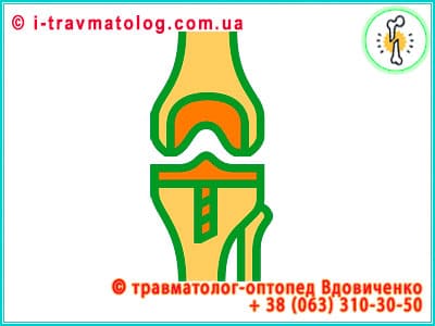
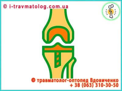
What is Knee Arthroplasty?
Knee joint arthroplasty (total arthroplasty, single-joint arthroplasty) in clinic No. 10 of the Institute of Orthopedics and Traumatology of the National Academy of Medical Sciences, Kyiv, Ukraine
The knee joint is one of the most sophisticated, biomechanically precise, and yet physiologically vulnerable mechanisms of the human body. As the largest synovial articulation, it daily accumulates and distributes colossal static and dynamic force vectors, providing us not only with elementary locomotion but also with the most complex coordinative stability. However, when the internal regenerative resource of tissues is finally exhausted, and the compensatory capacities of the ligamentous apparatus and subchondral bone are reduced to zero, the only effective solution becomes Knee Arthroplasty. This is a high-tech precision reconstruction involving the replacement of destructured biological components of the joint with innovative implants possessing the highest biocompatibility index.
Operation duration: 90-120 minutes.
Hospital stay after surgery is 7-14 days.
Today, this field of orthopedics and traumatology is undergoing a fundamental transformation: we are finally distancing ourselves from the simplified paradigm of "prosthesis as an inert spare part," moving toward a comprehensive interdisciplinary philosophy of functional restoration through the synergy of digital modeling, robot-assisted surgery, and breakthrough methods of cellular regeneration.
Etiology and Pathogenesis
The fundamental cause of systemic joint destruction is osteoarthritis (gonarthrosis). This is not merely mechanical wear of surfaces, but a complex molecular cascade of pathophysiological reactions. The process is characterized by progressive degradation of hyaline cartilage, pathological remodeling of the underlying (subchondral) bone, and chronic synovitis. When the friction coefficient increases and smooth gliding is replaced by agonizing "bone-on-bone" contact, irreversible tissue erosion is triggered.
- Degenerative-Dystrophic Factors: Genetically determined anomalies in type II collagen synthesis, age-related stagnation of chondrocyte proliferative activity, and critical disruption of microcirculation in epiphyseal zones. Over time, the extracellular matrix loses proteoglycans, loses hydrophilicity and elasticity, and becomes covered with a network of microcracks and fibrillations.
- Post-Traumatic Genesis: Consequences of severe intra-articular fractures of the condyles, invasive ruptures of the anterior cruciate ligament (ACL), and collateral structures, as well as iatrogenic consequences of total meniscus resection. Trauma fatally disrupts the biomechanical axis, creating zones of pathological hyperpressure where cartilaginous tissue undergoes necrosis in the shortest possible time.
- Immunopathological and Systemic Processes: Rheumatoid arthritis, psoriatic arthropathy, and other autoimmune nosologies. In these scenarios, the joint becomes a target for the aggressive impact of pro-inflammatory cytokines (TNF-alpha, interleukins), which induce the formation of pannus—an aggressive tissue that literally "devours" cartilage and bone.
- Metabolic and Endocrine Triggers: Morbid obesity, acting not only as a mechanical press but also as a source of adipokines—specific proteins of adipose tissue that maintain systemic inflammation. This also includes gouty arthropathy (microcrystalline lesion) and diabetes mellitus, which causes glycation of matrix proteins and deep ischemia of joint tissues.
Joint destruction is the result of a multifactorial cascade, where gonarthrosis and chronic inflammation lead to irreversible cartilage degradation, requiring radical restoration of the biomechanical axis.
Individual Reconstruction Strategy
Modern orthopedic science has completely moved away from unified templates. The choice of endoprosthesis configuration is based on a precision analysis of the degree of destruction of each of the three functional departments (compartments) of the knee: medial, lateral, and patellofemoral.
- Unicondylar (Partial) Knee Arthroplasty: Advisable for localized lesions of one department (usually medial). This is a highly selective, organ-preserving manipulation where only the defective segment is removed, while intact ligaments and healthy cartilaginous tissue are preserved. This ensures the preservation of proprioception—the natural sense of the joint in space.
- Total Knee Arthroplasty (TKA): Radical reconstruction of all articulating surfaces. The design includes a femoral component (cobalt-chrome alloys or oxidized zirconium), a tibial component (titanium alloys), and a high-tech insert made of ultra-high molecular weight polyethylene (UHMWPE), often enriched with vitamin E to minimize oxidation and wear processes.
- Revision Arthroplasty: The highest category of complexity, aimed at replacing previously implanted structures due to their wear, aseptic loosening, or periprosthetic infection. It requires the use of massive modular systems with intramedullary stems and metallic augments (blocks) to compensate for bone mass deficiency.
- Constrained (Hinged) and Posterior-Stabilized Systems (PS): Dictated by the necessity of severe valgus-varus deformation and ligamentous insufficiency. Such prostheses are equipped with a special mechanical "stabilizer" that takes over the role of the lost posterior cruciate ligament, guaranteeing the kinematic stability of the limb.
The choice of design varies from unicondylar systems to complex revision modules, where the key success factor is the precision selection of the individual implant and UHMWPE material for joint longevity.
Fundamental Diagnostics
Before intervention, the surgeon accumulates an exhaustive array of data for the verification of morphological deviations and the construction of a virtual surgical model:
- High-Resolution Weight-Bearing Radiography: Protocol study in an orthostatic position. It allows calculating the deviation angles of the mechanical axis of the limb with mathematical precision, assessing the state of "joint spaces," and identifying zones of subchondral sclerosis and osteophytosis.
- Magnetic Resonance Imaging (MRI): The gold standard for visualizing soft tissue structures. It allows detailed study of the state of patellar retainers, chondral defects, and the presence of bone marrow edema (BME), which is critical for predicting the outcome.
- Multispiral Computed Tomography (MSCT) with 3D Reconstruction: The foundation for preoperative digital planning. Tomography data are used to create patient-specific instruments (PSI) or to program robotic complexes ensuring implant positioning accuracy up to 0.5 mm.
- Comprehensive Laboratory Monitoring: Study of systemic markers (C-reactive protein, erythrocyte sedimentation rate, procalcitonin), as well as needle biopsy of synovial fluid if latent infection is suspected, which is an absolute contraindication to primary prosthesis installation.
The complex use of MRI, MSCT, and digital navigation systems allows for the creation of an ultra-precise architectural model of the intervention, minimizing the risk of component malposition.
Differential Diagnosis and Identification of "Red Flags"
Before making a final decision on invasive reconstruction, the physician must conduct a deep differential analysis to ensure that the source of suffering is indeed the knee joint and to exclude conditions that threaten life or require a completely different therapeutic vector.
- Radiating Pain (Referred Pain): Frequently, knee pain is a consequence of hip joint pathology (coxarthrosis) or degenerative processes in the lumbosacral spine (radicular syndrome L3-L4). A thorough physical examination with an assessment of the range of motion in adjacent segments and neurological status is mandatory.
- Vascular Pathology: Occlusive diseases of the lower limb arteries and deep vein thrombosis can mimic joint pain. A "red flag" here is the absence of pulsation in peripheral arteries, intermittent claudication, and leg edema.
- Infectious Status (Septic Arthritis): Acute onset, pronounced hyperthermia of the joint, fever, and sharp restriction of movement require immediate aspiration. Arthroplasty against a background of active infection is a fatal error leading to severe disability.
- Tumor Processes: Bone destruction can be caused by primary osteosarcomas or metastatic lesions. "Night pains" that do not resolve at rest, unmotivated weight loss, and atypical changes on images are direct indications for an oncological search.
- Neuropathic Pains: Complex Regional Pain Syndrome (CRPS) or diabetic neuropathy require conservative neurological treatment, as surgery in these cases may only intensify the pain syndrome.
Thorough screening for red flags, such as septic arthritis or referred pain, is necessary to exclude diagnostic errors and ensure the targeted nature of surgical treatment.
Protocol of Diagnostic Search and Exclusion of "Red Flags"
To ensure patient safety, a strict screening algorithm is implemented, aimed at identifying hidden contraindications and critical conditions.
- Stage 1: Clinical Verification of Pain and Neurological Screening. The physician conducts tests to differentiate the source of pain. Checking reflexes, foot muscle strength, and dermatome sensitivity excludes spinal canal stenosis and herniated discs.
- Stage 2: Exclusion of Ischemia and Venous Insufficiency. Mandatory ultrasound duplex scanning (USDS) of lower limb vessels. The presence of varicose veins or atherosclerotic plaques requires preliminary correction to prevent gangrene or fatal pulmonary embolism (PE).
- Stage 3: Sanitization of Chronic Infection Foci (Biological Safety). The patient undergoes an expanded examination by a dentist, otolaryngologist, and urologist. Any hidden infection is a risk of hematogenous spread of bacteria to the surface of the new implant, leading to its rejection.
- Stage 4: Endocrinological and Bone Audit. Assessment of glycated hemoglobin level (diabetes control) and bone tissue density (densitometry). Severe osteoporosis requires pharmacological preparation before prosthesis installation to prevent periprosthetic fractures.
An algorithmic approach to biological safety through infection sanitization and metabolic parameter control minimizes the risks of periprosthetic complications.
Surgical Technique
The implantation process is a synthesis of surgical art and strict engineering calculation aimed at restoring ideal biomechanics.
- Minimally Invasive Surgery (MIS): Use of specialized techniques (Subvastus or Midvastus approaches) that allow access to the joint without transecting the fibers of the quadriceps femoris muscle. This preserves its contractile ability and critically accelerates recovery.
- Precision Osteotomy: Use of oscillating saws and guide systems to excise affected areas. Bone surfaces are modeled to ensure maximum contact area with the prosthesis.
- Dynamic Soft Tissue Balancing: A critically important stage of adjusting ligament tension. The surgeon achieves symmetry of ligamentous balance in extension and flexion positions, which excludes implant laxity.
- Osteointegration Methods:
- Cemented Fixation: Use of bone cement (PMMA) for instant fixation.
- Cementless Fixation (Press-fit): Application of implants with a porous coating that stimulates the ingrowth of living bone tissue directly into the prosthesis structure.
The application of MIS technologies and the achievement of ideal ligament balancing are the keys to physiological load distribution and effective osteointegration.
Cellular Biotherapy
This vector represents an innovative convergence of classical orthopedics and molecular biology. We do not just change the mechanics; we modify the biological landscape of the joint.
- Platelet-Rich Plasma Therapy (PRP): Injection of the patient's own plasma with a high concentration of platelets. Released growth factors suppress chondrocyte death and arrest inflammation.
- Stromal Vascular Fraction Therapy (SVF): Extraction of multipotent cells from the patient's adipose tissue. These possess a powerful effect of "reprogramming" the joint's inflammatory environment into a regenerative one.
- Bone Marrow Aspirate Concentrate (BMAC): Contains a pool of stem cells and cytokines. Aimed at restoring bone tissue nutrition and stimulating the formation of cartilage-like tissue.
- Biomatrix Technologies and Autologous Chondrocyte Implantation (ACI): Use of biodegradable scaffolds seeded with the patient's own cells for "spot repair" of cartilage.
Using regenerative potential through stem cells and growth factors transforms the joint into an active biological environment, accelerating cellular restoration.
Regenerative Technologies and Biological Augmentation
The integration of innovative cellular therapy methods into the surgical protocol ensures optimal results at all stages of treatment.
- Hyaluronic Acid (HA): Acts as fundamental viscosupplementation. Before surgery, HA prepares the metabolic environment of the joint, reducing friction. After surgery, it improves the gliding of soft tissues and protects the UHMWPE insert from degradation.
- PRP (Platelet-Rich Plasma): Applied both before and during intervention to modulate the inflammatory response. Platelet growth factors activate neoangiogenesis and accelerate the healing of the surgical access.
- SVF and BMAC (Cellular Concentration): The role of these technologies is critical in cementless fixation. The introduction of SVF or BMAC into the "prosthesis-bone" contact zone provides explosive osteointegration, making implant fixation maximally reliable in the shortest time.
- ACI and MACI (Chondrocyte Transplantation): In partial joint lesions, MACI allows for the restoration of biological cartilage, which either postpones the date of total arthroplasty or is used in combination with it to restore intact joint sections.
- Combined Strategies: The combination of a matrix (HA), a cellular substrate (BMAC/SVF), and a biochemical stimulator (PRP) creates ideal conditions for prosthesis integration and reduces the patient's time on crutches due to an accelerated biological response.
The synergy of HA, PRP, and cellular concentrates (SVF/BMAC) creates a "biological bridge" for ultra-fast osteointegration and reduced rehabilitation periods.
Pharmacological Support and Nutritional Support
Successful prosthesis integration is inconceivable without pathogenetically substantiated drug therapy aimed at managing the body's systemic reactions.
- Anticoagulant and Antiplatelet Group: Vital drugs for preventing thromboembolic complications. Their effect lies in suppressing blood clotting factors and preventing platelet aggregation.
- Non-Steroidal Anti-Inflammatory Drugs (NSAIDs): Used to suppress inflammatory enzymes. They provide analgesia, reduce soft tissue edema, and promote early mobilization.
- Gastroprotector Group: Used in combination with anti-inflammatory therapy to protect the stomach and intestinal mucosa from damage.
- Broad-Spectrum Antibiotics Group: Perioperative prophylaxis aimed at creating bactericidal protection in joint tissues at the time of intervention.
- Drugs Affecting Bone Metabolism: Therapeutic effect aimed at increasing bone density for reliable endoprosthesis fixation.
- Angioprotectors and Venotonics: Contribute to improved microcirculation and venous return, accelerating hematoma resorption.
- Nutritional Support: Focus on amino acids, B vitamins, and antioxidants to stimulate collagen synthesis and regeneration of damaged nerve fibers.
Competent pharmacotherapy and anticoagulant protection form the necessary metabolic basis for arresting neurogenic inflammation and successful healing.
Recovery
The recovery process after total joint replacement is a complex biological cascade reaction requiring strict control at each stage.
- Acute Inflammation Phase (Days 1–5): Primary focus is on edema management and pain relief. The most important aspect is early verticalization, preventing stagnant phenomena.
- Early Proliferation Phase (Weeks 1–3): Active synthesis of granulation tissue begins. During this time, it is vital to maintain the elasticity of the joint capsule, preventing the formation of gross scar adhesions (arthrofibrosis).
- Remodeling and Adaptation Phase (Months 1–6): Period of final stabilization of prosthesis components and restructuring of the gait pattern, which was previously distorted by years of disease.
Sequential passage through the proliferation and remodeling phases under specialist control excludes arthrofibrosis and ensures adaptation to a new movement stereotype.
Rehabilitation
Rehabilitation after arthroplasty is a systemic neuromuscular retraining aimed at restoring proprioception and strength endurance.
- Kinesitherapy and Mechanotherapy: Use of passive trainers for continuous movement allows for gentle joint development. Subsequently, the focus shifts to strengthening the quadriceps femoris—the dynamic stabilizer of the prosthesis.
- Gait Pattern Restoration: Working with a rehabilitologist helps correct "habitual limping" and relearns the patient's correct body weight distribution.
- Physiotherapeutic Impact: Use of lymphatic drainage, electromyostimulation, and cryotherapy to accelerate effusion resorption and reduce disability periods.
- Psychological Adaptation: Overcoming the fear barrier of loading the operated leg and returning to active hobbies (swimming, cycling).
Integration of kinesitherapy and proprioceptive training methods restores dynamic limb control and returns the patient to full social activity.
Prevention and Long-Term Operation
The final result is 50% determined by the quality of postoperative follow-up.
- Early Kinesitherapy: Starting exercises in the first hours after awakening. Early weight-bearing prevents venous thrombosis.
- Continuous Passive Motion (CPM): Use of robotic mechanotherapy devices for gradual amplitude restoration.
- Postural Control and Proprioceptive Training: Teaching the patient a new walking scheme, posture correction, and strengthening stabilizer muscles.
- Long-Term Prevention: STRICT body weight control, exclusion of axial impact loads in favor of swimming or cycling. Mandatory annual radiological monitoring.
Compliance with the impact load protocol and regular radiological control are fundamental conditions for preventing aseptic loosening of the implant.
Patient Checklist
- Technical Basis: Which endoprosthesis model is chosen (material, fixation type) and why is it optimal for my bone morphology? Will digital navigation or individual PSI guides be used?
- Surgical Access: Is the use of Subvastus or Midvastus techniques planned to preserve the integrity of the quadriceps muscle?
- Biological Support: Which regenerative methods (PRP, BMAC, SVF) will be integrated into the surgery to accelerate osteointegration?
- Safety Protocol: What is the strategy for preventing thromboembolism and periprosthetic infection in my specific case?
- Prognosis: What is the expected range of motion and after what period can I return to full axial load?
An informed preoperative audit and selection of a minimally invasive tactic ensure patient psychological comfort and predictable functional output.
Nutritional Strategy for Regeneration
- Protein Foundation: Increasing the intake of high-quality protein (L-arginine, L-glutamine) to provide a substrate for soft tissue healing and bone integration.
- Micronutrient Stimulation: High doses of Vitamin C (collagen synthesis cofactor), Zinc (cell proliferation), and Vitamin D3 in combination with Vitamin K2 for directed calcium metabolism in the bone fixation zone.
- Antioxidant Protection: Omega-3 fatty acids in therapeutic dosages for modulating the inflammatory cascade and improving blood rheological properties.
- Neuroprotection: B vitamins for restoring the myelin sheaths of nerve endings affected during the surgical access.
Targeted nutritional support with microelements and amino acids acts as a catalyst for collagenogenesis and accelerates the structural restoration of the operated segment.
Clinical Research Results
- Osteointegration and Stability: Studies (Level I evidence protocols) demonstrate that the use of BMAC and SVF in cementless fixation increases the bone ingrowth index by 25-30% in the first 6 months. This significantly reduces the risk of early aseptic loosening.
- Pain Syndrome and Functionality: Patient groups receiving intraoperative PRP and HA show a decrease in VAS (Visual Analogue Scale) scores 40% faster in the early postoperative period compared to control groups. The WOMAC score improves by 15-20% above the average value by the end of the 3rd month.
- Rehabilitation Period: Using regenerative technologies allows for reducing the use of additional support means (crutches) by an average of 10-14 days due to accelerated soft tissue healing and reduction of secondary edema.
- Implant Longevity: Preliminary 10-year observation data indicate that optimizing the joint's biological environment with HA reduces UHMWPE wear by minimizing the abrasive impact of fibrin microparticles and inflammatory factors.
The application of evidence-based medicine methods confirms that cellular augmentation is a statistically significant factor in improving the functional survival of the joint.
Technology Synergy as the Key to Orthopedic Success
A deep analysis of the presented arthroplasty methodology allows for a definitive conclusion: modern orthopedics has reached a level where mechanical joint replacement has ceased to be an isolated operation, turning into a high-intelligence biological reconstruction system. The key to the optimal result lies in the integration of precision diagnostics (MRI, MSCT), robotic accuracy of implant installation, and powerful cellular support (PRP, SVF, BMAC). Such a combination allows not only for anatomical restoration but also for "reviving" surrounding tissues, ensuring the implant functions as naturally and durably as possible.
However, the effectiveness of all the above scientific achievements directly correlates with the time factor. Delaying consultation with a specialist in the presence of chronic pain syndrome leads to irreversible changes: severe muscle atrophy, bone base deformation, and degradation of neuromuscular connections, which significantly complicates both the surgery itself and the rehabilitation process.
Do not allow the depletion of your body's biological resource. Timely consultation with an orthopedic traumatologist at the stage of early clinical manifestations is not just a way to avoid pain, but a strategic opportunity to apply organ-preserving and regenerative methods, or to perform arthroplasty under conditions of preserved muscle tone and bone density. Only a fundamentally verified and timely approach guarantees a return to a completely active life with minimal risks and maximum functional results.
The harmonious combination of surgical precision and cellular technologies makes arthroplasty a highly reliable method, and timely diagnostics is the decisive link in achieving a durable result.
Summary
Knee arthroplasty in the era of high technology is not the final point in a medical history, but a powerful technological start that returns to the person the lost joy of full movement. The symbiosis of precision engineering thought in implant architecture with the colossal regenerative potential of cellular therapy allows us today to speak of a genuine "biological renaissance" of the joint. We provide patients with a quality of life that even a quarter-century ago was considered fantastic, turning mechanical replacement into a biological triumph.
Reference List
- Insall & Scott. Surgery of the Knee. 6th Edition. Elsevier, 2018.
- Berry D. J., Lieberman J. R. Surgery of the Hip and Knee: Principles and Practice. Wolters Kluwer, 2020.
- Filardo G., Kon E. et al. PRP Injections in Knee Osteoarthritis: What is the Evidence? Journal of Clinical Orthopaedics and Trauma, 2021.
- Hernigou P. et al. Cell Therapy for Osteoarthritis of the Knee. International Orthopaedics, 2019.
- Gobbi A. et al. BMAC and SVF in Regenerative Orthopaedics: A Systematic Review. Orthopaedic Journal of Sports Medicine, 2022.
- Canale S. T., Beaty J. H. Campbell's Operative Orthopaedics. 14th Edition. Elsevier, 2021.
- AAOS Clinical Practice Guideline. Management of Osteoarthritis of the Knee. Evidence-Based Guideline, 3rd Edition, 2022.
- Brittberg M., Gomoll A. H. Tissue Engineering and Cartilage Repair: ACI and MACI Procedures. The Journal of Bone and Joint Surgery, 2023.